From historic machinery in one of our own investigator’s personal collection to the modern technology transforming Wistar research today, microscopes have helped Wistar scientists and researchers around the world pursue innovative investigations.
A Curious Collection
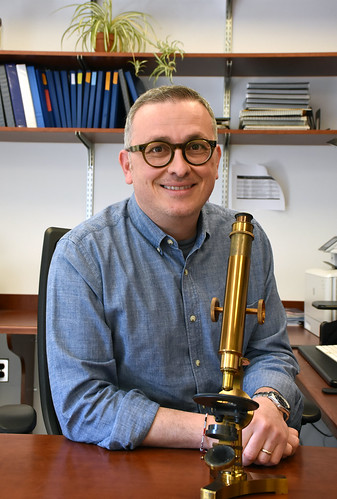
On a wooden desk sits 14 historical microscopes assorted in neat rows, gleaming a metallic gold in the office lighting. Manufactured between the 1800s-1900s in England and France, the instruments make up the personal collection of Dr. Italo Tempera, Associate Professor of Wistar’s Gene Expression & Regulation Program and Associate Director for Cancer Research Career Enhancement at Wistar’s Ellen and Ronald Caplan Cancer Center.
Tempera was interested in microscopy from a young age. “I started with biology when I was 13. I had a microscope at home,” he recalls. “I always wanted to collect and then at the beginning of the pandemic, I got the first one.” The microscope he refers to was made in 1830 in London and is the same kind of model that Darwin had. He also points out another model he finds most intriguing. Manufactured in France, the instrument was sold in Philadelphia from a shop on Chestnut Street, according to an inscription on the scope’s wooden box.
“We learn through our eyes. Microscopy is important because microscopes can reveal a lot of things we may not see through our experiments.” Tempera comments. “When you have a very powerful microscope image, you can really show and deliver your messages.” he continues.
The valuable knowledge that microscopes have given the scientific field – including how our cells work and are organized – emerged from these tools. In fact, Tempera and his colleagues recently used one from his collection (circa. 1890s – early 1900s London) to look at cancer tissue. “It was pretty good actually.” Tempera reveals in happy surprise.
The Horner Microscope
Tempera’s collection are not the only historical microscopes housed at The Wistar Institute. Nestled behind a glass display in the first floor Atrium is a microscope from 1830 made for Philadelphia anatomist William E. Horner. Made by Austrian optical instrument maker Simon Plossl, this microscope is still functional today. In 2017, Wistar’s Imaging Facility Managing Director James Hayden used the Horner microscope to image tumor cells and reconstructed skin from Wistar labs. He imaged the same samples with a modern Nikon microscope in Wistar’s Imaging Facility and compared the results.
“One of the things that surprised me was just how good it was. The fact that you could still separate cells and you could still see nuclei means that people 150 years ago could see the same stuff we can see now.” Hayden shares. “Even though technology has marched ahead, the basics haven’t changed much.”
What has changed with advances in microscopy is how images are used in scientific research. “The images are not just documentary anymore. They can be quantified. They become the data.” Hayden emphasizes.
Modern Microscopy at Wistar
Now many types of advanced microscopes exist, enhanced by the innovative technology of today’s world. From confocal microscopy to electron microscopy, the ability to magnify and quantify what would normally go unseen has expanded. Wistar research is helped tremendously by such powerful microscope technology at the Imaging Facility. For example, the Facility has imaged drug treatments on melanoma cells, stitched tissue images together, and recorded live cell behavior over mere milliseconds.
As a Shared Resource, Wistar’s Imaging Facility impacts diverse realms of research both internal and external to the Institute. “We have biological questions that need to be answered, so we take our existing technology, and we work with our industry partners to come up with new ways of imaging something to bypass the problem.” Hayden describes.
Whether it is virology or anatomy, biochemistry or chemical engineering, microscopy can be applied. Hayden says that it is the “investment in a facility that can keep up with the technology and more importantly, the hiring of people that can continually train others to support research” that makes microscopy at The Wistar Institute special.