Author: The Wistar Institute

Wistar Science Synergy Through Fostering International Collaborations

Inaugural graduate students from Leiden University Medical Center speak to their Wistar experience.

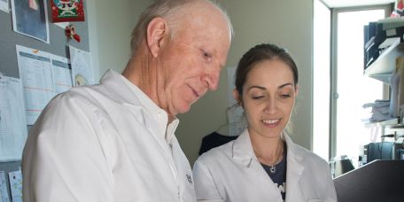
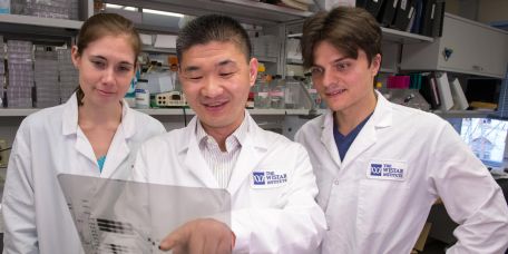
The Wistar-Schoemaker International Postdoctoral Fellowship is a special exchange of postdoctoral fellows between our two institutes. We sat down with Katarina Madunić and Tamas Pongracz to hear more about this important science interchange and what is next in their research careers

What is your scientific expertise and how is it connected to Wistar?

Katarina Madunić: My mentor Dr. Manfred Wuhrer got to know Dr. Mohamed Abdel-Mohsen during a collaborative seminar that we organized together with Wistar and LUMC to exchange current research results. We learned very early on that we have mutual interests and might benefit from each other’s expertise. Dr. Abdel-Mohsen’s work is focused on various aspects of HIV biology and glycoimmunology. My Ph.D. was in cancer research where I studied glycosylation of colorectal cancer – specifically a largely unexplored type of glycosylation called mucin type O-glycosylation.

In my research at Leiden University Medical Center (LUMC), I discovered that sugar molecules attached to proteins (glycans) are specifically expressed by tumors, yet never appear in normal tissue of the patient. This makes glycans promising targets for antibody-based immunotherapy. At LUMC, our focus is on structural glycomics where we explore which glycans are expressed in different tissues and cells in different diseases. This is complementary to Dr. Abdel-Mohsen’s lab, which has more of a focus on the role of different glycans in the immune system, and in diseases like HIV.

In HIV infection, there is an interplay between gut tissue, its glycosylation, and microbiota. Translocation of the microbiota during HIV infection causes long term inflammation leading to health complications. The key question this work is trying to answer is whether glycosylation of the gut cells is linked to the diversity of the microbiota and their translocation during HIV infection. At Wistar, I am exposed to different techniques such as lectin array and mass spectrometry to analyze gut glycosylation. My expertise is in mass spectrometry, so when I observed the data, I brought a different perspective to its interpretation.

Tamas Pongracz: Our antibodies – crucial in the fight against the SARS-CoV-2 virus – are coated with sugars in a process called glycosylation. Part of my Ph.D. research focused on antibody glycosylation signatures in COVID-19 infection. Using mass spectrometry, I identified these antibody-linked sugars and found specific coating patterns that are associated with disease severity at hospitalization. Based on these patterns, we could predict how a patient’s disease would progress. The study took place within the framework of a multi-disciplinary collaboration at LUMC, turning the pandemic into a scientifically fruitful period. This is a complementary time to be at Wistar and observing the research taking place Mohamed Abdel Mohsen’s lab. Interestingly, sugars can modify their inflammatory potential depending on how antibodies are coated —Mohamed’s lab specializes in this topic. During my time, I got to see how specific sugars affect antibody function

We are so grateful to be here and supported through this Fellowship that brings researchers from Leiden to Wistar to nurture new scientific collaborations. While we are here, the visit also has a diplomatic flavor because we are the first visitors from Leiden. We are pioneering the collaboration between the two labs.

What are some next steps for you both – scientifically and professionally?

KM: I accepted a postdoctoral fellowship in Copenhagen, Denmark on the team of Dr. Adnan Halim. There, my work will be more focused on understanding what specific function glycans have on a particular protein. My long-term plan is to come back to the Netherlands after the postdoctoral fellowship. Colon glycosylation and it’s interplay with the gut microbiota in the context of different inflammatory diseases as well as cancer is a subject that sparks my interest the most, and I would like to continue working on this subject in the future.

TP: Once we are back at Leiden, both of us will spread the news about how great Wistar is and that international collaboration is key for both institutes. I feel very much attached to glycobiology. In the future, I would like to be part of an interdisciplinary team studying the mechanistic aspects of glycosylation, a field currently gaining more and more attention.

Anything to add about your experience?

KM: Dr. Abdel-Mohsen and members of his lab welcomed us very warmly here. Samson, Ferlina, and Shalini gave us a thorough understanding of the assays they utilize. They also organized a lab night out where we went bowling and got to know each other better.

TP: Leadership has welcomed us so warmly. We dined with Anne Schoemaker—her late husband Hubert is who this Fellowship has been named after, and we heard how supportive and brilliant he was as a scientist and how human he was. That was touching, and we are very privileged to be supported by this Program. I think that this has been a great kickstart of the collaboration. We already have ideas on what to explore when we return home and it’s going to be fruitful for everyone.

Discussing the Mysteries of Melanoma

A Q&A with Drs. Chengyu Liang and Meenhard Herlyn on skin cancer research at The Wistar Institute, including a few questions from melanoma survivors.

Melanoma is the most aggressive form of skin cancer. Work at The Wistar Institute’s Melanoma Research Center aims to understand the biology behind the disease to help develop new therapies and improve existing treatments. In this Q&A, we spoke with Dr. Chengyu Liang, a professor in the Molecular & Cellular Oncogenesis Program at Wistar’s Ellen and Ronald Caplan Cancer Center who joined Wistar in 2020 and Dr. Meenhard Herlyn, director of The Wistar Institute Melanoma Research Center and Professor in the Molecular & Cellular Oncogenesis Program. The research they conduct aims to have impacts on melanoma patients and survivors. Thus, this National Melanoma and Skin Cancer Awareness Month, we included questions from melanoma survivors and staunch Wistar supporters Eleanor Armstrong and Pat Dean who organize an annual fundraiser walk for the Institute’s melanoma research program.

Dr. Liang, what attracted you to studying melanoma?

Dr. Chengyu Liang: My passion and curiosity! A few years ago, we identified a so-called “sunscreen” gene that helps cells to repair after they have been damaged by UV rays; and we know that UV-induced DNA damage is a major risk factor for melanoma – the most aggressive type of skin cancer. This inspired us to find out more about how this genetic sunscreen system plays in human beings and what it means to melanocytes (the origin of melanoma) and melanoma when the system is running out of order.

What are you working on now specifically regarding melanoma and what is this work’s potential impact on cancer treatments?

CL: The question that we are always intrigued by is: What makes melanoma melanoma? Compared to most other types of human cancers, one striking feature of melanoma is that its genome is flooded with mutations associated with UV-induced damage. Now, the burning questions are: What drives such genetic change? What are the molecular mechanisms underlying the UV-footprint in melanoma? Understanding these mechanisms not only enable early-risk prediction but also help oncologists and researchers to develop cancer treatments with responses that have long-term durability.

Second, melanoma originates from an easily spread and multipotent cell population, which can help explain the inborn aggressiveness and treatment resistance of the skin cancer. In addition to targeting specific pro-cancer mechanisms to kill cancer cells that are often, if not always, encountered by tumor resistance, is it possible to force cancer cells to resume the process of normal growth control or differentiation? To this end, we are trying to understand the molecular mechanisms that control melanoma differentiation. This work holds promise to identify new vulnerabilities in melanoma that can be targeted to revert the negative effects of mutations and strengthen anti-tumor immune responses to melanoma.

How important is a person’s immune system in preventing skin cancer and is there anything that can be done to strengthen a person’s immune system against melanoma?

CL: It can’t be more important! Our immune system is like the ‘shepherd’; its duty is to keep ‘sheep’ protected. Melanoma is like the ‘wolf’. A powered immune system is an experienced shepherd that could easily and quickly identify and target the wolf in the flock and clear it up. As a matter of fact, what immunotherapy does is release the built-in brake system of our immune system and revive and direct its killing energy to cancer cells.

The immune system is an ecosystem – a complex network of cells, tissues, and organs that orchestrate to maintain homeostasis to protect human bodies against internal and/or external assaults. If a well-balanced immune system is considered healthy to life, a well-balanced life would also be considered beneficial to the immune system. Many tips have been suggested such as healthy diet, exercise, mental and physical support, etc. Notably, fasting and calorie restriction (CR), have been shown experimentally and in clinical trials, to be able to slow and even stop the progression of cancer, kill cancer cells, boost the immune system, and significantly improve the effectiveness of chemotherapy and radiation therapy. With our growing understanding of this sophisticated system, we might be able to come up with more effective strategy to boost it or manipulate it to outsmart cancer.

You’ve previously done research on UV radiation causing mutations that contribute to melanoma. What are some daytime hours that have the highest risk of damaging UV radiation and sun exposure?

CL: This depends. The lighter the skin, the more sensitive to sunburn and/or skin damage by UV rays. Human skin pigmentation is an evolutionary adaptation to UV radiation. Fair skinned people may easily burn within 20 minutes of exposure to direct sunlight, thus wearing a broad-spectrum sunscreen is always encouraged. Certainly, you also want to have a strong genetic sunscreen system as noted before.

What future directions will you take your work?

CL: Our ultimate goal is to make melanoma a curable disease. The current challenge is that we still have more than 50% of melanoma patients who are not responsive to any treatment. We need to solve this puzzle. We also need to figure out what’s the right therapy to be used for the right patient at the right time. Why does therapy work in some patients, but not others? We believe that prevention is the most effective treatment of melanoma. Thus, identifying a new biomarker for early detection is equally important and urgent. All these challenges depend on a deeper understanding of the biology of melanoma as a cancer, and the biology of melanoma interaction with the human body as a system.

Dr. Herlyn, why is building the cancer research talent at Wistar important (eg. Dr. Chengyu Liang joining the Cancer Center)?

Dr. Meenhard Herlyn: Every research field needs a critical mass of researchers, both junior and senior. Any institution needs ‘new blood’, meaning investigators who may have experience, knowledge, and connections in the critical field. Dr. Liang brings important expertise not only to Wistar but the entire larger field here and beyond. Her work on DNA damage and repair fills an important gap for us.

What would you like to see for the future of melanoma research at Wistar?

MH: Melanoma has been a major focus for research at Wistar. We have developed a strong outreach program in which we collaborate not only with our colleagues at Penn but also with numerous national and international research laboratories. Melanoma research at Wistar should remain multi-disciplinary. Our strong biology is the foundation and platform for collaborations that ensure continuing progress. Melanoma investigators should incorporate cutting edge technologies and strategies. We have developed strong ties to our clinical colleagues, and we expect that in the future these ties will strengthen as our research more and more directly benefits patients. Thus, we not only translate research from bedside-to-bench but also from bench-to-bedside.

Why are supporters such as those fundraising and donating to the Institute important?

MH: There are many areas of research that are essential to run a successful program but that cannot be funded through National Institute of Health or industry grants. For example, there are meetings with other scientists to exchange ideas and develop new collaborative strategies, seed funding for new projects that are still in the exploratory phase, collaborations with clinicians for specimens and preparing reagents for diagnostic studies, developing high risk/high gain projects and ideas, and obtaining new research tools. Supporters like our donors and fundraisers are integral to our work at Wistar, without whom we could not have as great an impact as we do.

Spotlighting Wistar’s Cheyney University Student Scientists

Read some reflections from the Cheyney University students who participated in Wistar’s Biomedical Research Methods course during Spring 2022.

Lights, lab coats, action! The click of pipettes and low murmur of voices fill the open lab space as students set up experiments. Each undergrad is seated at their own lab station equipped with a row of pipettes, colorful tube holders, and other tools they need for today’s lesson. It is one of the final weeks of Wistar’s Biomedical Research Methods course, taught by Dr. Kristy Shuda McGuire, The Wistar Institute dean of Biomedical Studies, and Dr. Ian Tietjen, research assistant professor in Wistar’s Montaner Lab.

This educational collaboration between The Wistar Institute and Cheyney University engages STEM undergraduates in a hands-on laboratory experience focused on practical bench skills including cutting-edge biomedical research techniques. The students are working on real Wistar science with a focus on cancer biology. The curriculum uses a project based on Wistar research from the laboratory Dr. Maureen Murphy, program leader, Molecular & Cellular Oncogenesis Program, Ellen and Ronald Caplan Cancer Center, which centers on African-centric variants of the p53 tumor suppressor gene—the most frequently mutated gene in cancer. Once students successfully complete the course, they have opportunities to continue conducting research through summer internships at Wistar.

Below are some students’ reflections of the BRM course.

“I got to do some of the things here that we only spoke about in virtual lab, so actually pouring the agar plates was really cool. I really liked that, and it was so fun.”

– Sophia Kurian, sophomore. She is studying biology with an interest in pursuing a career involving forensic science and plans to continue research at Wistar through the summer internship program.

“I took this course because I wanted to get a research background. It’s a very fun environment and a program that really showed me practical applications of science. It made me realize science is a lot more of an art. … My favorite skill to learn was definitely working with cell lines in the cell culture lab. I loved using the aspirator.”

– Abdiel Mandella Reynolds, sophomore. He is studying biology with a pre-medicine focus and plans to go to med school. He plans to continue research at Wistar through the summer internship program.

“I picked Wistar’s course because of the hands-on experience. I started school during the pandemic, and it was so different. But here, we get that in-person, hands-on experience, that allows us a broader understanding of science in a lab.”

– Michelle Lucas, sophomore. She is studying biology and her current career goal is to be a travel nurse.

“This course really makes you think about how people use certain tools or mechanisms for all these different applications. It shows you how to use different devices and use your knowledge in a lab. I heard that researchers from UPenn come over here to use some of these really advanced tools which is something I’m happy that I’m around.”

– Mohamedanwar Idress, sophomore. He is studying biology with a pre-medicine focus and plans to go to med school.

Wistar’s Shark Tank Pitch Competition Goes Swimmingly

The hybrid event marked the conclusion of the Institute’s Life Science Innovation Course that provides experience in biotechnology entrepreneurship.

Students enrolled in The Wistar Institute’s Life Science Innovation course entered the shark tank. They shrugged off nerves, put their best selves forward, and made mock business pitches to a panel of expert biotechnology investors. The students had been preparing their pitch in teams based off technology leveraging Wistar intellectual property and interacted with inventor scientists and industry experts to design solutions to development and commercialization issues.

The competition included pitches from Wistar predoctoral students, Wistar postdocs, Wi-Star interns, and undergraduate students from Cheyney University and La Salle University. Pitch topics covered unique biotechnology and scientific discoveries with clinical potential such as HIV and pregnancy biomarkers and diagnostic testing for ovarian cancer and COVID-19.

The panel of judges included Anthony Green, Ph.D., chief scientific officer of Ben Franklin Technology Partners; Donna Cordner, cofounder and managing partner of OKM Capital; and Tom Penn, partner of MVP Capital Partners. After each pitch, the judges evaluated the teams and discussed detailed breakdowns of investment plans, appropriate funding requests, important legal distinctions around Intellectual Property, and establishing an appropriate market model.

“The Shark Tank event is the culmination of months of effort by teams of undergraduate students and Wistar trainees to develop compelling business strategies for Wistar technologies. By examining the complexity of technology commercialization, participants have expanded their understanding of translational science and will hopefully be inspired to apply an entrepreneurial lens to their own research.” says David Zuzga, Ph.D, associate dean of Biomedical Studies.

A reception followed the competition. Please see the album below for photos of the event. 

Life Sciences Innovation Shark Tank Event 2022

Wistar Receives PAsmart Grant to On-Ramp More Apprentices from Our Biomedical Education & Training Programs 

For more than 20 years, The Wistar Institute has been training community college students with relevant, much-needed skills to attain in-demand jobs in the biomedical and biotechnology fields. Since 2000, Wistar understood that biomedical lab technicians would always be highly sought after for their own labs and beyond, and thus set out to create a program to cultivate this much-needed talent.

That program became the flagship Biomedical Technician Training Program, now a registered pre-apprenticeship program. The opportunity to extend hands-on training allowed Wistar to develop the Biomedical Research Technician Apprenticeship—the first registered, non-traditional apprenticeship in biomedical research in the nation.

Upon being awarded a PAsmart grant of $649,551 from the Pennsylvania Department of Labor & Industry, Wistar is now on track to expand both programs further across the region and create additional pre-apprenticeships to offer more students the opportunity to prepare for careers with additional employers in the growing life sciences industry.

Workforce Development at Wistar 

In this virtual event held by the Chamber of Commerce, Wistar’s Associate Dean of Biomedical Studies Dr. David Zuzga spoke about workforce development and the value of apprenticeship programs at the Chamber of Commerce virtual event “Finding Emerging Talent in STEM”.

On April 20, 2022, the Chamber of Commerce for Greater Philadelphia hosted a virtual event reviewing how to address challenges in recruiting and training a STEM workforce. Guest speakers included our Associate Dean of Biomedical Studies Dr. David Zuzga who spoke about strengthening the STEM workforce through education and apprenticeship opportunities such as Wistar’s Biotechnician Training (BTT) Program.

Attracting and retaining STEM talent in the workforce, especially in an increasingly informationally complex and interdisciplinary world, has been a challenge faced by employers and educational institutions alike. As Philadelphia grows into a leading life sciences hub, developing and retaining STEM talent is at the forefront of workforce development initiatives.

Zuzga commented on the advantages of an integrated approach to traditional education with workforce training. He highlighted Wistar’s BTT program which collaborates with Community College of Philadelphia to provide a laboratory orientation boot camp where participants work on authentic Wistar research projects and are then matched with summer internships in academic and industry settings.

“There is a transformation in the program’s participants. It’s remarkable to be so connected to an employer-partner and provide that type of value in a nontraditional program.” said Zuzga. He continues, “It’s essential for employers to begin engaging with educational institutions to better inform the type of competencies they are looking for in a successful workforce.” Zuzga explains that as employers collaborate with training providers to address lab skills that are more specific and relevant, they are creating more value for participants as they receive training more transferable to STEM careers.

Wistar Scientists Link Mutation to a Severe Lung Disease

Newly published research identifies a mutation associated with scarring of the lungs, revealing a useful diagnostic tool and target for gene therapy.

Idiopathic pulmonary fibrosis (IPF) is a fatal lung disease that can cause death within five years of diagnosis. In an international research collaboration recently published in the Journal of Experimental Medicine, Wistar scientists and colleagues in the Netherlands identified a mutation on telomeres – which protect the ends of chromosomes – that helps explain this disease. This study directly links the telomeric protein POT1 (Protection of Telomeres Protein 1) to IPF.

Emmanuel Skordalakes, Ph.D., Associate Professor in the Gene Expression and Regulation Program at Wistar and senior author of the paper, previously published research in Nature Communications that delved into the structure and function of POT1, the protein that binds the ends of our chromosomes (telomeres) and protects them from DNA damage response (DDR).

In this new study, Skordalakes and his team used patient data obtained by their Dutch counterparts and conducted a series of biophysical and biochemical experiments to understand how telomeres with mutated POT1 are linked to IPF. Proteins usually have particular sites, which allows them to bind to molecules that match that specific pocket. The team observed that in this chronic lung disease, the mutated POT1 affected how the protein binds to the telomere and thus its ability to prevent DDR.

“We found the mutated POT1 does not bind the telomere as efficiently as the wild type one,” said Skordalakes. The less effective binding of POT1 leaves telomeres exposed to DDR. When this occurs, cells undergo a process called senescence that stops them from functioning and dividing.

“We looked at the [cells of] these patients and they are senescent,” Skordalakes shares. Senescent lung cells build up and contribute to many symptoms of IPF like difficulty in breathing.

The POT1 mutation will be added to a medical database to be used in hospitals to help diagnose patients with IPF as well as provide doctors a target for gene therapy. Skordalakes says, “When you know that your work has an impact on patients directly in the clinic – that’s what’s exciting.”

Wistar Scientists Reveal New Hypothesis in How Epstein-Barr Virus Hijacks a Host

Newly published research identifies proteins in EBV-infected cells that decreased expression of genes linked to the spread of the virus, a fresh direction for EBV research.

Epstein-Barr virus is a common, long-lasting virus that affects most individuals. While those infected are often asymptomatic, the virus has been linked to various immunocompromising diseases and cancers—popular news recently reported on a strong connection to Multiple sclerosis (MS). Published in PLOS Pathogens, a new collaborative study led by Italo Tempera, Ph.D., revealed how lamins – proteins in the nucleus of a cell – could affect EBV replication in host cells by influencing the virus genome, ultimately changing how virus genes function.

When a virus such as EBV infects cells, it invades the nucleus of host cells to replicate and spread. Previous research by Tempera – Associate Professor of The Wistar Institute Gene Expression & Regulation Program and Associate Director for Cancer Research Career Enhancement at Wistar’s Ellen and Ronald Caplan Cancer Center – showed that changes to the virus genome helps EBV sustain its infection in hosts.

“Viruses don’t reinvent but use existing machinery,” says Tempera. “EBV has evolved to take every mechanism the host uses to regulate its presence in the nucleus, and it has such profound consequences for the genome that are really fascinating.”

To investigate how genes are regulated to lead to this persistent infection, Tempera and colleagues infected and imaged genetically altered B cells, a type of immune cell. They also employed sequencing to study the virus genome. The team found that lamins – specifically lamins A/C – repressed gene expression linked to replication of the virus’s genetic material.

The team also observed that the infection of B cells with EBV produced the same effect as the activation of B cells by T cells in an immune response. This copying of B cell biology by the virus may help explain why EBV is latent (not recognized by the immune system right away) and further clarify how host cells respond to the invasion of EBV into the nucleus.

“It’s a very basic mechanism,” he says, “but one that can give us a lot of good ideas about how the virus can hijack the host. It’s a really new angle.”

Tempera hopes this work will open new directions in which to study viruses and inspire other researchers to explore how viruses interact with hosts. Regarding therapeutic potential, he muses that with more research, scientists might be able to identify targets for EBV infection that could prevent or turn off genetic expression in cancer. “That could be the long haul of how you move this basic science into something more practical.”

A Closer Look into Our Scientific Enterprise

Dr. Luis Montaner, Wistar’s Associate Director for Shared Resources, and Mark Drinker, Wistar’s Associate Director for Administration of the Ellen and Ronald Caplan Cancer Center, chat about core facilities that support the innovations of our Wistar scientists, particularly spotlighting Wistar’s new Humanized Models of Disease Core.

Q: Can you tell our readers why it is so important that Wistar has Shared Resources? We’re a small biomedical research institute, why do we need it?

Mark: Wistar Shared Resources are mission critical. Every institution needs to have core facilities that support and are tailored to the science taking place in their organizations or institutes. Shared Resources allow faculty to incorporate leading-edge technology into their science in order to advance their research programs. Shared Resources may be created as a result of technology developed within an individual laboratory. For example, a laboratory may develop technology and expertise that is critical to advance their own science, and over time many other labs find they have that same need. This could be the impetus of the technology to be scaled into a Shared Resource designed to meet the needs of many researchers.

Q: Can you explain your work as the Associate Director for Administration of the Ellen and Ronald Caplan Cancer Center Shared Resources?

Mark: I work closely with Dr. Montaner, our Shared Resources director, and our Cancer Center leadership to position our core facilities as research engines that serve as extensions of our faculty laboratories. The goal is to maintain state-of-the-art Shared Resources that advance the research efforts of Wistar faculty and research partners. To accomplish this, our Shared Resources all contain technology, high-tech equipment, and exceptional personnel who have the expertise to work with each individual laboratory at Wistar.

Q: Can you describe some of the goals of the new Humanized Models of Disease Core, as its Scientific Director?

Luis: With the new Core under Dr. Zhe Yuan as Managing Director, we hope to generate a resource of humanized mice that could be of service to infectious disease scientists within the Vaccine and Immunotherapy Center, as well as the Cancer Center faculty. This model allows researchers to study how tumors from persons grow or respond to therapy if implanted in mice as well as how the human immune cells interact with tumors. The core is an investment to generate enough expertise in-house so we can bring a humanized mouse (differentiated with the human immune system) to the laboratory. The core will allow Wistar teams to utilize the humanized mouse as needed to move their experiments forward.

Q: What are humanized models of disease and why are they important?

Luis: A humanized model of disease is utilizing an animal model to study human physiology. The ability to study human immune cells within tissue environments for long periods of time (weeks) provides for studies that may better resemble our human body. We can use these models to study the infectious disease process of pathogens that infect human blood cells such as HIV. We can also use it for understanding human tumors.

Q: What was the impetus behind creating the Humanized Models of Disease Core Facility?

Mark: For many years Wistar faculty, including Drs. Herlyn and Montaner, have been developing mouse models to study cancer and infectious disease. Our scientists need to utilize models that closely replicate the human immune system to test their hypotheses. By creating a Shared Resource, we hope to develop a structure that allows Wistar to maintain this promising technology and offer it broadly to our research teams in order to advance their science.

Luis: Innovative science is made possible by the strong support of our donors. This new facility is just one example of the progress Wistar can accomplish with the generosity from our philanthropic community. The ability to propel research forward is facilitated by those who believe Wistar science is an impactful investment for the future. The creation of the new center is supported by a philanthropic commitment to Wistar’s most important priorities, which is to advance our technologies and scientific resources available to our researchers. The discoveries that will be made are the fruits of investing philanthropically in The Wistar Institute.

Q: Can you give me some examples of the scientific information that can be collected from using these models?

Luis: To study novel therapeutic strategies, you can use a tissue culture plate that just has the cells that you want to interact or understand. But once you start reconstituting the way our bodies are, then you must start scaling up into systems too. How well can I reach the blood with this drug that is in circulation to maintain this change? How well can I add a drug that would reach the cells in a physiological state? If it’s an organ system, how well can I get to that organ for my treatment to still work in the microenvironment of the lung, or the liver, or the gut? As we look for what model systems do, we must start scaling into a whole system.

When it comes to generating a platform to pilot test some proof of principle or evidence that this treatment could work in the context of human cell interactions, the humanized mice model becomes one tool that is a lot less costly and a lot less demanding with respect to using limited resources. The use of the humanized mouse model becomes a tool that could potentially give a researcher more confidence that what they’re working on is worth advancing.

Q: Can you describe some of your own HIV research and how it could be impacted by this new facility?

Luis: The humanized mouse model gives you the ability to screen many ideas within a model. Underway in our BEAT-HIV Martin Delaney Collaboratory efforts at Wistar, we are developing and testing novel strategies of viral eradication by 1) activating the immune system to clear HIV-infected cells, and 2) directly targeting HIV by CRISPR CAS9 and other gene editing approaches to rid the viral infection directly from the cells. This model gives us the ability to screen multiple strategies that either target infected cells directly or target the immune system to clear infected cells due to the immunotherapy.

Q: What do you hope that researchers will get out of using this facility?

Luis: As the technology keeps moving forward, the application of humanized mouse models in cancer research are starting to become a higher priority. For example, we can place patient tumors into mice that have compatible immune systems matched with the tumor. It’s also going to be important to test immunotherapies or chemotherapies in the context of cancer eradication as well as for the infectious disease research of the Vaccine and Immunotherapy Center.

We all have a particular signature to our immune systems, and the best model is to generate a mouse that has the same signature of the person that the tumor has been growing in (i.e., creating a personalized research model). Studying therapy approaches where a tumor also matches the immune system of the person in the same mouse allows us to identify personalized cancer therapies. These types of models are now becoming more possible.

Zooming in on Microscopes at Wistar

From historic machinery in one of our own investigator’s personal collection to the modern technology transforming Wistar research today, microscopes have helped Wistar scientists and researchers around the world pursue innovative investigations.

A Curious Collection

On a wooden desk sits 14 historical microscopes assorted in neat rows, gleaming a metallic gold in the office lighting. Manufactured between the 1800s-1900s in England and France, the instruments make up the personal collection of Dr. Italo Tempera, Associate Professor of Wistar’s Gene Expression & Regulation Program and Associate Director for Cancer Research Career Enhancement at Wistar’s Ellen and Ronald Caplan Cancer Center.

Tempera was interested in microscopy from a young age. “I started with biology when I was 13. I had a microscope at home,” he recalls. “I always wanted to collect and then at the beginning of the pandemic, I got the first one.” The microscope he refers to was made in 1830 in London and is the same kind of model that Darwin had. He also points out another model he finds most intriguing. Manufactured in France, the instrument was sold in Philadelphia from a shop on Chestnut Street, according to an inscription on the scope’s wooden box.

“We learn through our eyes. Microscopy is important because microscopes can reveal a lot of things we may not see through our experiments.” Tempera comments. “When you have a very powerful microscope image, you can really show and deliver your messages.” he continues.

The valuable knowledge that microscopes have given the scientific field – including how our cells work and are organized – emerged from these tools. In fact, Tempera and his colleagues recently used one from his collection (circa. 1890s – early 1900s London) to look at cancer tissue. “It was pretty good actually.” Tempera reveals in happy surprise.

The Horner Microscope

Tempera’s collection are not the only historical microscopes housed at The Wistar Institute. Nestled behind a glass display in the first floor Atrium is a microscope from 1830 made for Philadelphia anatomist William E. Horner. Made by Austrian optical instrument maker Simon Plossl, this microscope is still functional today. In 2017, Wistar’s Imaging Facility Managing Director James Hayden used the Horner microscope to image tumor cells and reconstructed skin from Wistar labs. He imaged the same samples with a modern Nikon microscope in Wistar’s Imaging Facility and compared the results.

“One of the things that surprised me was just how good it was. The fact that you could still separate cells and you could still see nuclei means that people 150 years ago could see the same stuff we can see now.” Hayden shares. “Even though technology has marched ahead, the basics haven’t changed much.”

What has changed with advances in microscopy is how images are used in scientific research. “The images are not just documentary anymore. They can be quantified. They become the data.” Hayden emphasizes.

Modern Microscopy at Wistar

Now many types of advanced microscopes exist, enhanced by the innovative technology of today’s world. From confocal microscopy to electron microscopy, the ability to magnify and quantify what would normally go unseen has expanded. Wistar research is helped tremendously by such powerful microscope technology at the Imaging Facility. For example, the Facility has imaged drug treatments on melanoma cells, stitched tissue images together, and recorded live cell behavior over mere milliseconds.

As a Shared Resource, Wistar’s Imaging Facility impacts diverse realms of research both internal and external to the Institute. “We have biological questions that need to be answered, so we take our existing technology, and we work with our industry partners to come up with new ways of imaging something to bypass the problem.” Hayden describes.

Whether it is virology or anatomy, biochemistry or chemical engineering, microscopy can be applied. Hayden says that it is the “investment in a facility that can keep up with the technology and more importantly, the hiring of people that can continually train others to support research” that makes microscopy at The Wistar Institute special.

Microscopes at Wistar